Pedicle morphology of the first sacral vertebra
- Location: Çankaya, Ankara, Turkey

Okutan O, Kaptanoglu E, Solaroglu I, Beskonakli E, Tekdemir I.
Posterior transpedicular screw fixation has been widely used for the management of unstable lumbosacral spine caused by trauma, degenerative conditions, congenital defects and neoplasms. Knowledge of the pedicle diameters of the first sacral vertebra is crucial for safe placement of the screws. Thirty dry sacral specimens (18 male, 12 female) were used for study of the first sacral pedicles. Cephalad-caudad height, anterior-posterior width, transverse and sagittal angles, and depth of S1 pedicle were presented. The mean width of the pedicles were estimated as 22.5±2.6 mm and 22.2±2.8 mm; the heights were 13.6±2.3 mm and 13.6±2.7 mm; the depths were 50.7±3.7 mm and 51.8±3.5 mm for female and male, respectively. The mean transverse angles were 43°±2.3 and 41°±2.2; the sagittal angles were 19°±2.9 and 19°±3.7 for female and male, respectively. The depth and the angle of screw trajectory is as important as entrance point for pedicular screw placement to the S1 to avoid injury to the vascular structures anteriorly and nerve roots medially. © Neuroanatomy. 2003; 2: 16-19.

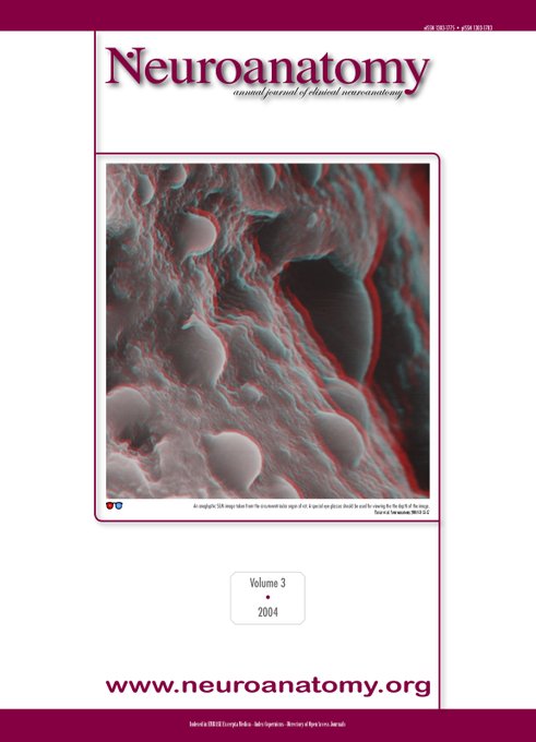
Neuroanatomy № 15 | http://neuroanatomy.org/15
Related articles
-
 A case of bilateral high division of the sciatic nerves, together with a unilateral unusual course oFreeVolume 2 [2003] Çankaya (Ankara) 03/01/2023Mas N, Ozeksi P, Ozdemir B, Kapakin S, Sargon MF, Celik HH, Yener N. In a 62-year-old male cadaver, high division of the sciatic nerve was observed bilaterally. Additionally, on the right side of the same cadaver, the common peroneal nerve passed jus...
 -
 A case of progressive multifocal leukoencephalopathy (PML): diffusion - weighted MR imaging findingsFreeVolume 2 [2003] Çankaya (Ankara) 03/01/2023Oguz B, Karli Oguz K, Akpinar E, Cila A, Sain Guven G. Progressive multifocal leukoencephalopathy (PML) is a progressive subacute demyelinating disease caused by neurotropic papova virus, usually in immunocompromised patients. As the number of cases ...
 -
 A simple low-cost method for two dimensional microscopic measuring and stepping on the microscopic pFreeVolume 2 [2003] Çankaya (Ankara) 03/01/2023Adiguzel E, Duzcan SE, Akdogan I, Tufan AC. In this study, a simple low cost method to be used in morphometric studies on microscopic anatomical structures is described. Increasing need for stereological methods depend on laboratories equipped with s...


Useful information
- The landing page for NEUROANATOMY is neuroanatomy.org.
- Instructions to authors page is To authors
- You can submit your article via this email address editor.neuroanatomy@gmail.com
- You can also use our EndNote style.
Comments
Leave your comment (spam and offensive messages will be removed)